
Figure 1.
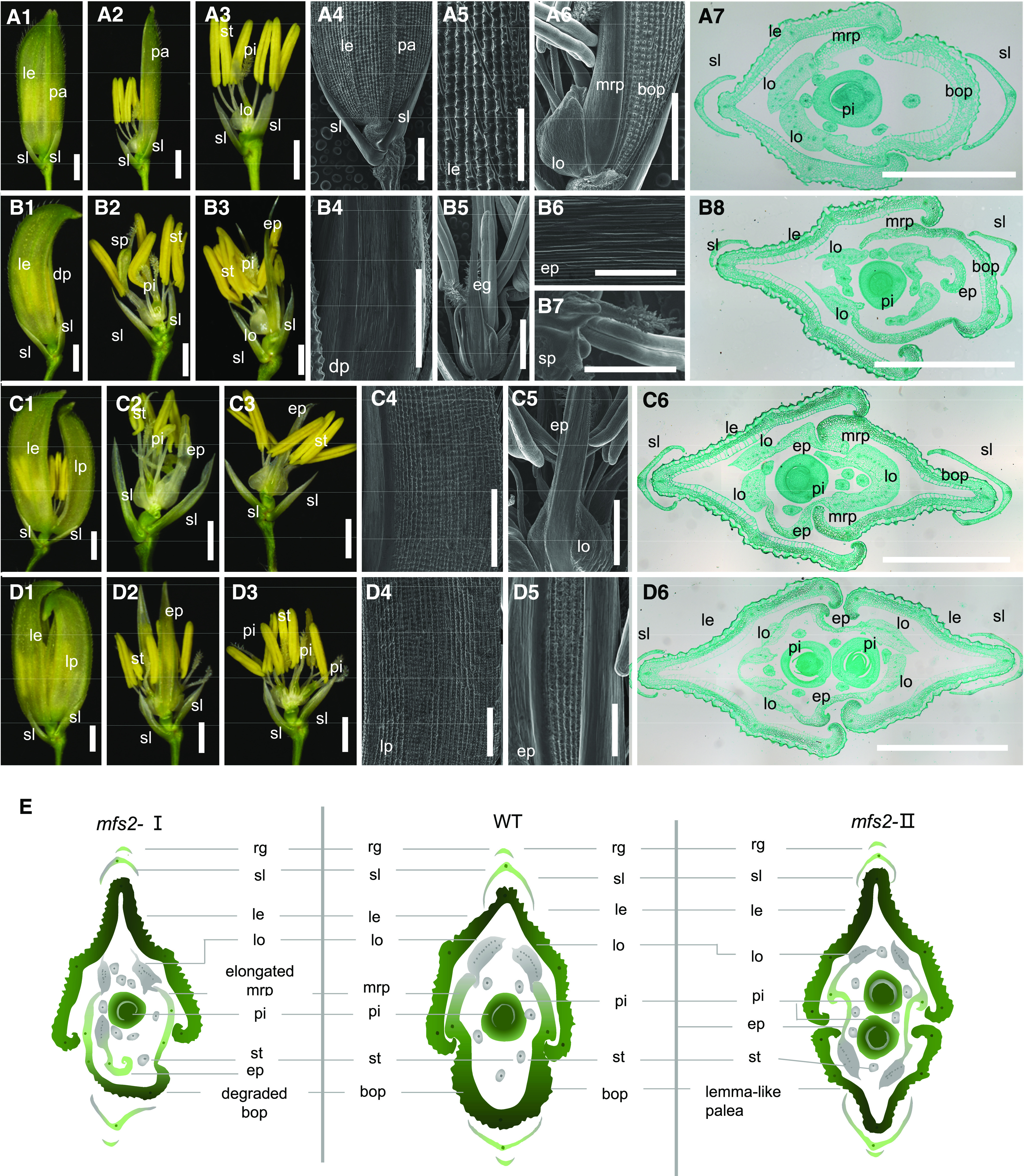
Phenotypes of spikelets of the wild-type and mfs2. A1, Complete spikelet of the wild type. A2, Spikelet from A1with lemma (le) removed. A3, Spikelet from A1 with lemma and palea (pa) removed. A4, Surface of a wild-type spikelet. A5 and A6, Surface characters of le, lodicule (lo), bop, and mrp, respectively. A7, Transverse sections of a wild-type spikelet. B1, mfs2 spikelet with degraded palea (dp). B2 and B3, Spikelet with lemma and palea removed. B4 and B5, Surfaces of the degraded palea and extra glume, respectively. B6, Magnification of B5. B7, Surface of the stamen-pistil fusion organ (sp). B8, Transverse sections of a mfs2 spikelet with degraded palea. C1, mfs2 spikelet with a partially lemma-like palea (lp). C2 and C3, Spikelet with lemma and palea removed. C4 and C5, Surfaces of the partially lemma-like palea and extra glume, respectively. C6, Transverse sections of a mfs2 spikelet with a partially lemma-like palea. D1, mfs2 spikelet with a lemma-like palea. D2, Spikelet from D1 with lemma and lemma-like palea removed. D3, Spikelet from D2 with the extra palea/mrp-like organ removed. D4 and D5, Surfaces of the lemma-like palea and extra glume, respectively. D6, Transverse sections of a mfs2 spikelet with lemma-like palea. E, Schematic diagrams of wild-type (WT) and mfs2 spikelet structure in transverse section. ep, Extra palea/mrp-like organ; pi, pistil; sl, sterile lemma; st, stamen. Scale bars = 1 mm (A1–A3, B1–B3, C1–C3, and D1– D3), 200 μm (A4–A6, B4– B7, C4, C5, D4, and D5), and 500 μm (A7, B8, C6, and D6).