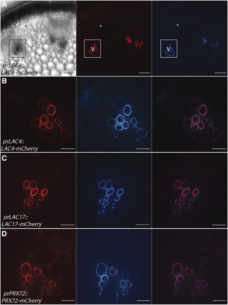
Figure 2.
AtLAC4-mCherry, AtLAC17-mCherry, and AtPRX72-mCherry localize to the SCW of xylem vessel elements during stage 1. Representative images depicting brightfield, the tagged LAC or PRX under its respective native promoter (red), UV lignin autofluorescence (blue), and merged image are shown. V, Xylem vessel element. Asterisks indicate cells that will differentiate into interfascicular fibers. A, Low magnification of AtLAC4-mCherry provides context for localization. Outlined region is vascular bundle with early lignifying xylem vessel elements. B, High magnification of AtLAC4-mCherry in the SCW of xylem vessel elements. C, High magnification of AtLAC17-mCherry in the SCW of xylem vessel elements. D, High magnification of AtPRX72-mCherry in the SCW of xylem vessel elements. Scale bars = 50 µm (A) and 20 µm (B–D).

