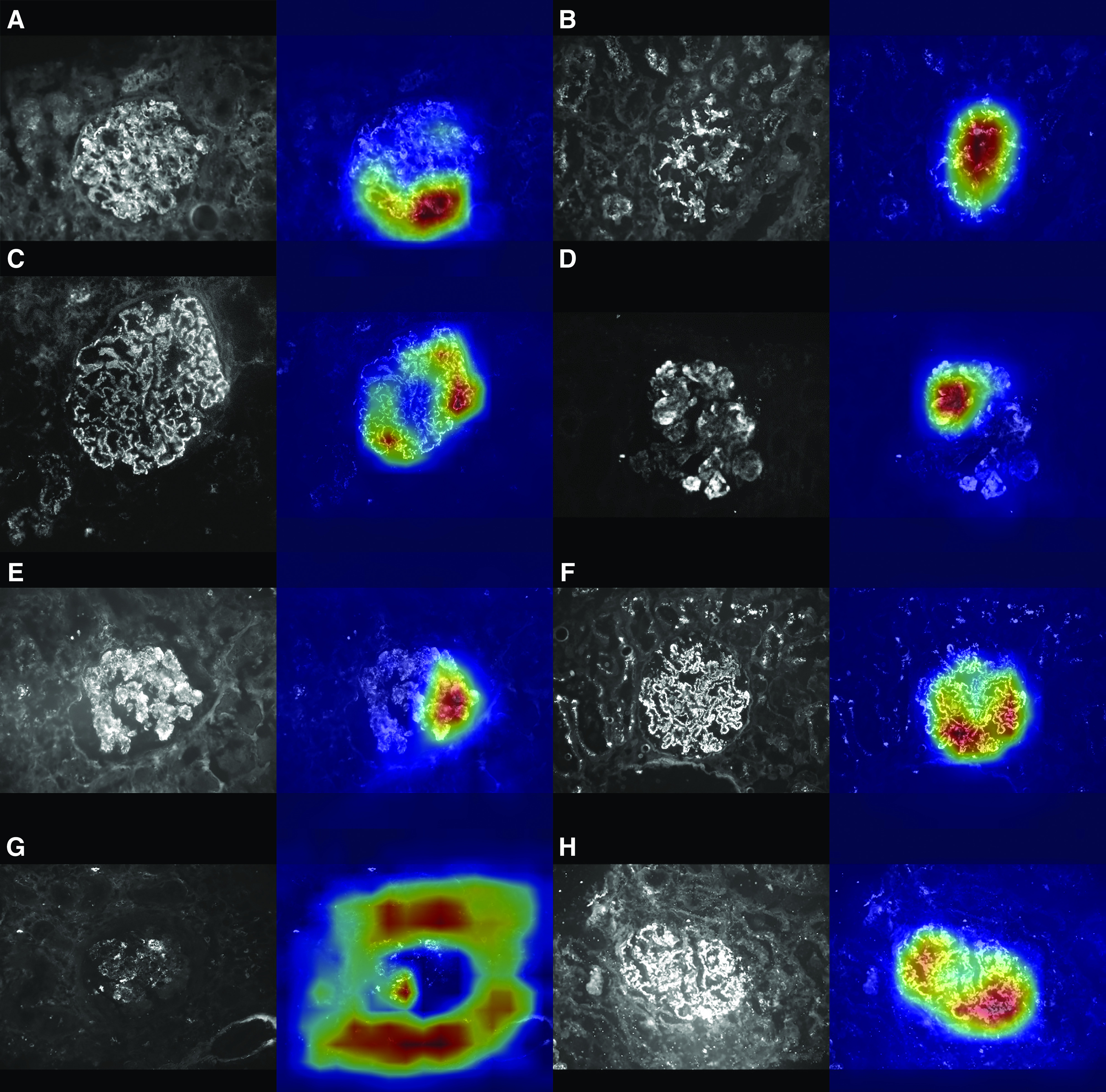
Figure 3.

The Convolutional Neural Network classification is influenced by the deposits on the glomerular structure inside the image. Each panel of the figure represents the result of the feature-specific convolutional neural networks elaboration of a test image. In (A–H), left panels show the original test images, while right panels show their heat maps. In the heat map, the red area shows the sections of the image most involved in the classification process by the convolutional neural network. All of the presented images were correctly classified by the specific convolutional neural network: (A) parietal, (B) mesangial, (C) continuous regular capillary wall, (D) irregular capillary wall, (E) coarse granular, (F) fine granular, (G) segmental, (H) global.
