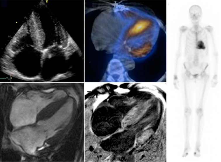
Fig. (5).
This is an example of multimodality imaging of a patient with TTR-CA and aortic stenosis. The echocardiogram (top left) demonstrates LVH. Cardiac scintigraphy (right) demonstrates cardiac amyloidosis which is more obvious via single-photon emission on CT (upper middle). Cardiovascular MRI with and without gadolinium enhancement on the bottom. Treibel et al. (A higher resolution / colour version of this figure is available in the electronic copy of the article).

