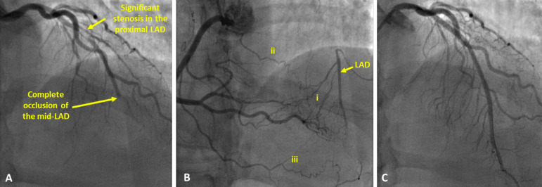
Fig. (1).
Angiography images of a patient with a Chronic Total Occlusion (CTO) of the Left Anterior Descending artery (LAD). (A): contrast injected into the left coronary artery demonstrates severe stenosis in the proximal part of the LAD, contrast flow stops abruptly at the point of complete occlusion in the mid-LAD. (B): contrast injected into the right coronary demonstrating retrograde filling of the LAD via multiple collateral channels, both septal (i) as well as epicardial (ii, iii) channels can be identified. (C): contrast injected into the left coronary artery after successful revascularisation of the LAD. Two stents have been implanted (one in the proximal LAD and a second at the site of the occlusion). (A higher resolution / colour version of this figure is available in the electronic copy of the article).

