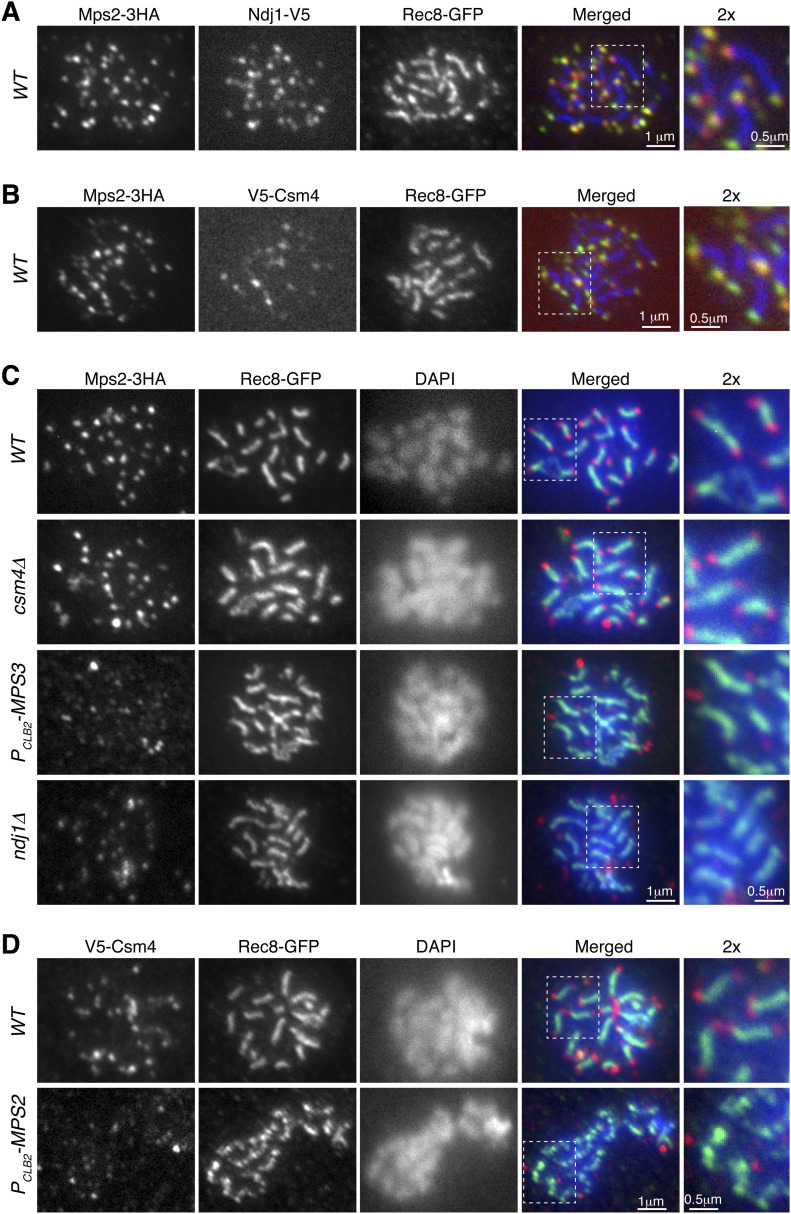
Figure 3. Mps2 is a telomere-associated protein.
Meiotic cells were harvested for nuclear spreads, followed by immunofluorescence to probe V5-, HA-, and GFP-tagged proteins. DAPI stains DNA. Rec8 is used to mark the chromosome axis. Enlarged views (2×) are shown to the right. (A) Representative cell showing colocalization of Mps2 with Ndj1 to meiotic telomeres. Red, Ndj1-V5; green, Mps2-3HA; blue, Rec8-GFP. (B) Representative cell showing colocalization of Mps2 and Csm4 at telomeres. Red, V5-Csm4; green, Mps2-3HA; blue, Rec8-GFP. (C) Representative cells showing that telomeric localization of Mps2 depends on Mps3 and Ndj1 but not Csm4. Red, Mps2-3HA; green, Rec8-GFP; blue, DAPI. Note that Mps2 localizes to the chromosome ends in WT and csm4Δ cells. (D) Representative cells showing telomeric localization of Csm4 depends on Mps2. Note that chromosome axes appear less compacted in the PCLB2-MPS2 cell. Red, V5-Csm4; green, Rec8-GFP; blue, DAPI.

