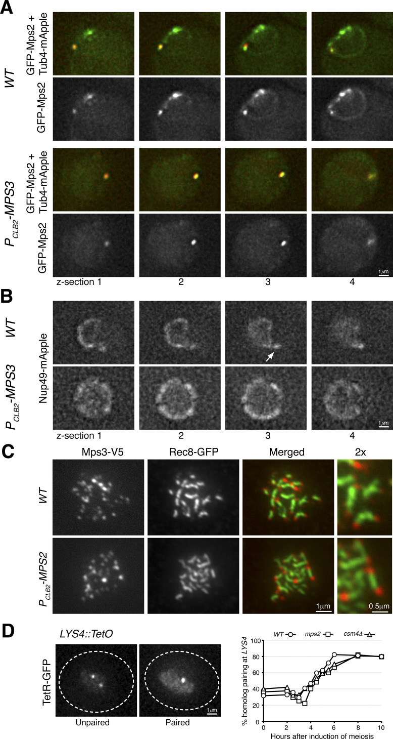
Figure S2. Mps2 localization and homolog pairing in meiosis.
(A) Live-cell fluorescence microscopy showing GFP-Mps2 (green) localization at prophase I. Note that GFP-Mps2 is clustered around the nuclear periphery in the wild-type (WT) cell but is only visible at the spindle pole body in the PCLB2-MPS3 cell. Four continuous optical sections are shown. Tub4-mApple (red) marks the spindle pole body. (B) Live-cell fluorescence microscopy showing Nup49-mApple localization at prophase I. The arrow points to the nuclear protrusion in the wild-type cell. Four continuous optical sections are shown. (C) Meiotic Mps3 binds to telomeres. Surface nuclear spreads were prepared as in Fig 3. Note that Mps3 remains bound to chromosome ends in the Mps2-depleted cell. Rec8 marks the chromosome axis. Red, Mps3-V5; green, Rec8-GFP. (D) Quantification of homolog pairing at the LYS4 locus. Cells were induced to undergo synchronous meiosis; aliquots were withdrawn at indicated times. TetR-GFP forms a nuclear focus when chromosome IV homologs are paired at LYS4. At least 100 cells were counted at each time point. Three biological replicates were performed; one representative is shown.

