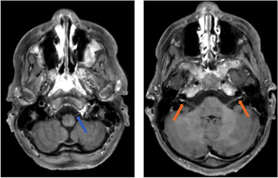
FIGURE 1.

Axial slices of brain magnetic resonance imagery (MRI) (T1w TSE SPIR images). Cranial nerves enhancement upon gadolinium injection is shown: blue arrow pointing the hypoglossal nerve (left panel) and the two red arrows pointing the two facial nerves (right panel)
