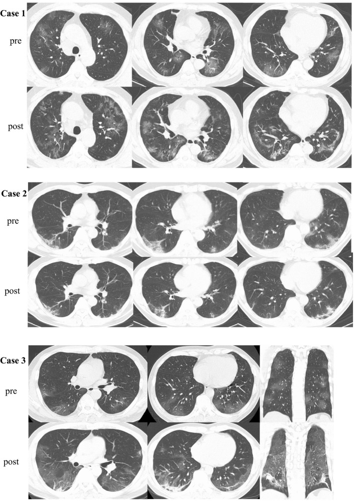
FIGURE 2.

Comparison of CT findings between pre‐ and post‐treatment for COVID‐19 pneumonia. In case 1, the post‐treatment chest CT revealed that GGO in bilateral lungs were increased, furthermore intra‐ and interlobular septal thickening region, bronchiectasis within GGO were emerged. In case 2, the chest CT images reveal bilateral small ground‐glass opacities with linear shadows located dorsally in the lower lung lobes. Post‐treatment chest CT revealed while a part of GGO regions were improved improvement, the linear shadow in GGO was thickened. In case 3, bilateral GGO and septal thickening region were progressed. The regions were predominantly in dorsal. Abbreviation: CT, computed tomography; COVID‐19, coronavirus disease 2019; GGO, ground‐glass opacities
