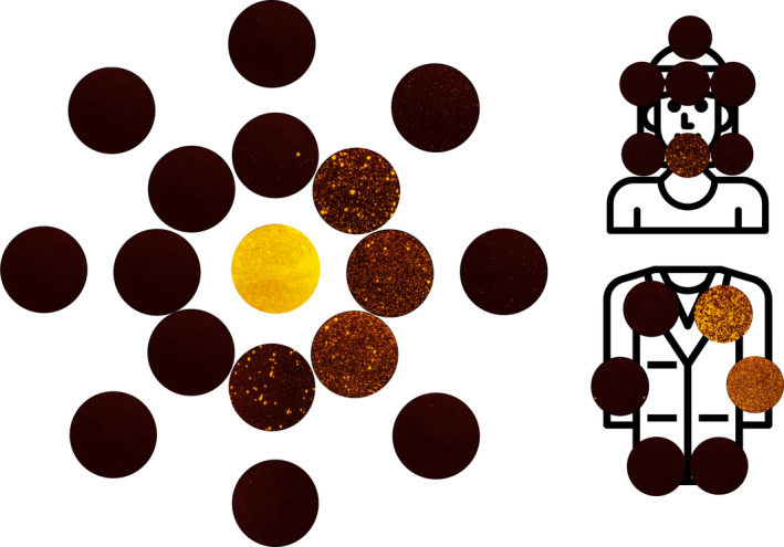
Figure 5.

Composite image comprised of all filter paper samples images used for image analysis within the first 1 m from the one repetition of the anterior crown preparation (no suction) condition. Colour balance and contrast adjusted to aid visualisation. Samples are arranged with the central sample in the centre, and samples from 0.5 m and 1 m arranged concentrically moving outwards. The axis is the same as demonstrated in Figures 1, 2, 3, 4 [Colour figure can be viewed at wileyonlinelibrary.com]
