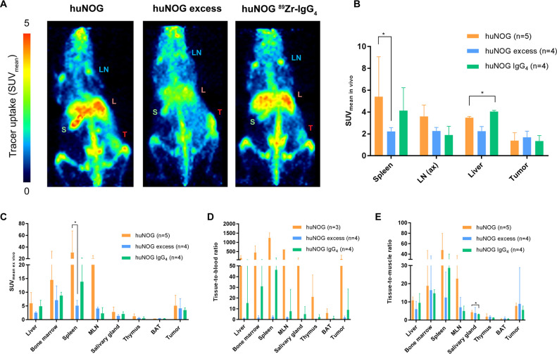
Figure 1.
In vivo PET imaging and ex vivo biodistribution of 89Zr-pembrolizumab in immunocompetent humanized NOG mice. Mice were xenografted with A375M tumor cells and received tracer injection at day 0. For blocking studies huNOG mice received a 10-fold excess of unlabeled pembrolizumab (huNOG excess). As a control for non-specific uptake huNOG mice were injected with 89Zr-IgG4. PET imaging performed on day 7 post injection (pi). (A) In vivo PET examples (maximum intensity projections) at day 7 pi showing uptake in tumor (T), axillary lymph nodes (LN), liver (L) and spleen (S). (B) In vivo uptake of 89Zr-pembrolizumab in spleen, lymph nodes (axillary), liver and tumor, at day 7 pi. Uptake is expressed as SUVmean. (C) Ex vivo biodistribution of 89Zr-pembrolizumab in humanized NOG mice. Uptake is expressed as mean radioactivity per gram tissue, adjusted for total body weight (SUVmean ex vivo). Data expressed as median±IQR *p≤0.05. BAT, brown adipose tissue; huNOG, humanized NOG mice; MLN, mesenteric lymph nodes; PET, positron emission tomography.

