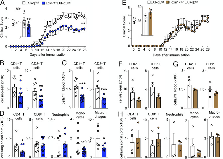
Figure 7.
T cell LXRαβ deficiency protects mice from autoimmune disease. (A–C) After EAE induction in 2-mo-old LXRαβfl/fl versus Lck-LXRαβ−/− mice, clinical score, with inset representing area under curve (AUC) quantified for each mouse (A) and frequency of splenic (B) and circulating (C) CD4+ T cells and CD8+ T cells at day 28 after EAE. (D) Frequency of spinal cord CD4+ T cells, CD8+ T cells, neutrophils, monocytes, and macrophages at day 28 after EAE. (A–D) Data are n = 8–10 per group from two independent experiments. *P < 0.05, **P < 0.01, ***P < 0.001. (E–G) After EAE induction in 2-mo-old LXRαβfl/fl versus FoxN1-LXRαβ−/− mice, clinical score with inset representing AUC for each mouse (E; n = 5 per group) and frequency of splenic (F) and circulating (G) CD4+ T cells and CD8+ T cells at day 28 after EAE (n = 3 or 4 per group). (H) Frequency of spinal cord CD4+ T cells, CD8+ T cells, neutrophils, monocytes, and macrophages at day 28 after EAE. (E–H) Data are n = 3–5 per group and are representative of two independent experiments. Statistical analysis used was Student’s t test (two groups only) or two-way ANOVA followed by Bonferroni’s post-test (two groups over time). All data are mean ± SEM.

