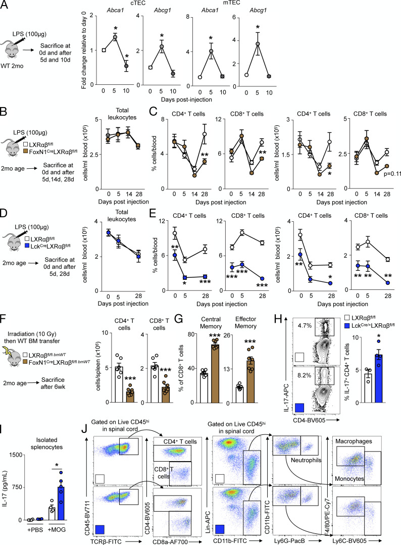
Figure S5.
LXRαβ is required for efficient thymic recovery, reconstitution of the T cell pool, and EAE. (A) Abca1 and Abcg1 levels by real-time PCR from FACS-sorted WT cTECs and mTECs over time after LPS injection. Data are n = 3 or 4 group and are representative of two independent experiments. *P < 0.05. (B) In 2-mo-old LXRαβfl/fl and FoxN1-LXRαβ−/− mice, mice were injected with LPS (100 µg) intraperitoneally and sacrificed 5, 14, or 28 d after injection, and circulating leukocytes were enumerated after LPS injection. (C) Frequency and enumeration of circulating CD4+ and CD8+ T cell levels after LPS injection are also shown. (D) In 2-mo-old LXRαβfl/fl and Lck-LXRαβ−/− mice, mice were injected with LPS (100 µg) intraperitoneally and sacrificed 5, 14, or 28 d after injection; circulating leukocytes were enumerated after LPS injection. (E) Frequency and enumeration of circulating CD4+ and CD8+ T cell levels after LPS injection are also shown. (B–E) Data are n = 4–7 per group from two independent experiments. *P < 0.05, **P < 0.01, ***P < 0.001. (F) In 2-mo-old LXRαβfl/fl and FoxN1-LXRαβ−/− mice, mice were lethally irradiated and transplanted with WT BM and sacrificed after 6 wk, with circulating CD4+ and CD8+ T cell levels enumerated. (G) Quantification of frequency of effector memory and immunoinhibitory splenic CD8+ T cells is also shown. (F and G) Data are n = 6 or 7 per group from two independent experiments. ***P < 0.001. (H) Representative flow cytometry plots and quantification of IL-17+CD4+ T cells after 5-d Th17 polarization and 4-h PMA/ionomycin/GolgiPlug stimulation ex vivo from naive 2-mo-old LXRαβfl/fl and Lck-LXRαβ−/− mice. Data are n = 3 or 4 per group and are representative of two independent experiments. *P < 0.05. (I) IL-17 concentration (measured by ELISA) from supernatants of splenocytes stimulated with MOG35–55 peptide ex vivo for 3 d from EAE-treated 2-mo-old LXRαβfl/fl and Lck-LXRαβ−/− mice. Data are n = 4 or 5 per group and are representative of two independent experiments. *P < 0.05. (J) Representative flow cytometry plots of spinal cord gating of leukocytes from EAE-treated 2-mo-old LXRαβfl/fl and Lck-LXRαβ−/− mice. Lin, lineage (CD3, CD90.2 CD19, NK1.1). Statistical analysis used was Student’s t test or two-way ANOVA followed by Bonferroni’s post-test. All data are mean ± SEM.

