Figure 1.
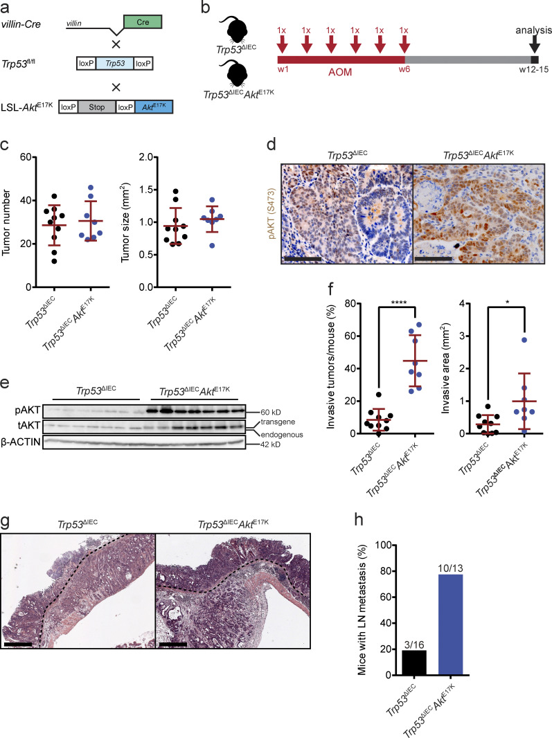
AOM-treated Trp53ΔIECAktE17K mice develop highly invasive tumors with lymph node metastasis. (a) Schematic representation of the strategy used for the generation of Trp53ΔIECAktE17K mice (fl, floxed; wt, wild type). (b) Experimental design of the AOM treatment of Trp53ΔIECAktE17K mice. (c) Primary tumor number and primary tumor size in AOM-treated Trp53ΔIEC and Trp53ΔIECAktE17K mice. Data are mean ± SD, n ≥ 8 per genotype. Not significant by t test. (d) Representative images of pAKT immunohistochemistry (IHC) on sections from AOM-induced colon tumors in Trp53ΔIEC and Trp53ΔIECAktE17K mice. Scale bars = 100 µm. (e) Immunoblot analysis of phosphorylated (p) and total (t) AKT levels in whole-tumor lysates of AOM-treated Trp53ΔIEC and Trp53ΔIECAktE17K mice. Lower band corresponds to endogenous AKT; upper band represents transgenic AKT. β-ACTIN was used as loading control. n = 7 per genotype. (f) Frequency of invasive tumors per mouse and extent of invasive area in AOM-treated Trp53ΔIEC and Trp53ΔIECAktE17K mice. Data are mean ± SD, n ≥ 8 per genotype; *, P < 0.05; ****, P < 0.0001 by t test. (g) Representative images of H&E-stained tumor sections from AOM-treated Trp53ΔIEC and Trp53ΔIECAktE17K mice (dashed lines mark invasive fronts). Scale bars = 500 µm. (h) Frequency of mice with lymph node metastasis in AOM-treated Trp53ΔIEC and Trp53ΔIECAktE17K mice. n ≥ 13 per genotype. Numbers indicate the mice with metastasis/all mice.

