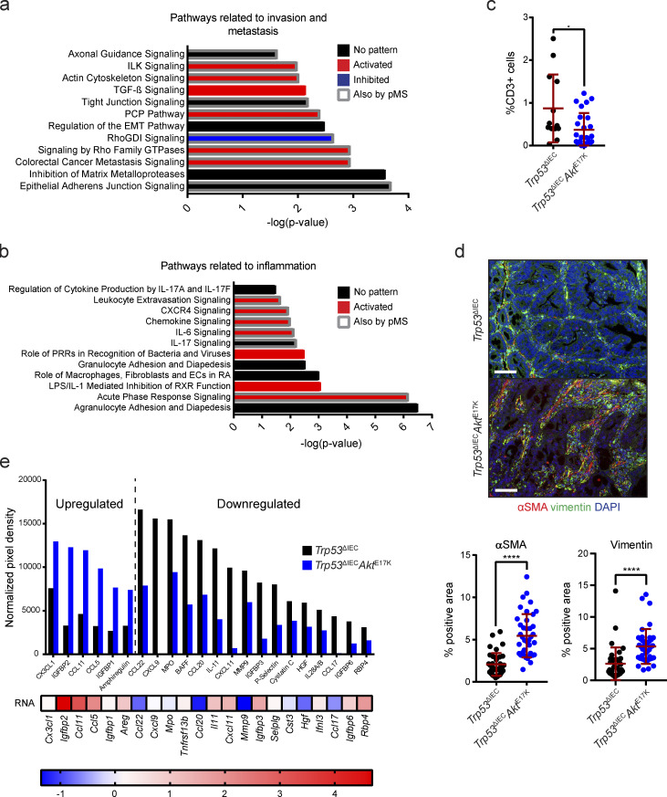
Figure S1.
Transcriptomic and phosphoproteomic analysis of Trp53ΔIECAktE17K tumors reveals regulation of pathways associated with invasion and metastasis as well as inflammation. (a and b) Signaling pathways associated with invasion and metastasis (a) as well as inflammation (b), which are significantly regulated in response to AKT hyperactivation. Predictions were made based on RNA sequencing of whole tumor tissues from AOM-treated Trp53ΔIEC and Trp53ΔIECAktE17K mice using IPA. P < 0.05 with right-tailed Fisher’s exact test, activation: z-score > 0, inhibition: z-score < 0, no pattern: pathway is differentially regulated, but z-score is not available. Regulated pathways which were also identified by phosphoprotein-specific MS of whole-tumor tissues of AOM-treated Trp53ΔIEC and Trp53ΔIECAktE17K mice using IPA are highlighted by a gray frame. P < 0.05. (c) Quantification of CD3 immunohistochemistry (IHC) in the invasive tumors of Trp53ΔIEC and Trp53ΔIECAktE17K mice. Cell numbers are shown as percentage of total cells, and at least one tumor and one invasive front was quantified per mouse. Data are mean ± SD; *, P ≤ 0.05 by t test. ROUT test with Q = 1% was used to remove outliers. n = 4 mice per genotype. (d) Representative images and quantification of immunofluorescent analysis of αSMA (red) and vimentin (green) expression in the tumors of Trp53ΔIEC and Trp53ΔIECAktE17K mice. Cell nuclei are stained with DAPI (blue). Data are mean ± SD; ****, P ≤ 0.0001 by t test. n ≥ 3 mice per genotype. Scale bars = 100 µm. (e) Cytokine array analysis of whole-tumor lysates from Trp53ΔIEC and Trp53ΔIECAktE17K tumors. A protein was considered to be up-regulated in Trp53ΔIECAktE17K tumors with a fold change >1.5 and was considered to be down-regulated with a fold change <0.75. n ≥ 6 per genotype.

