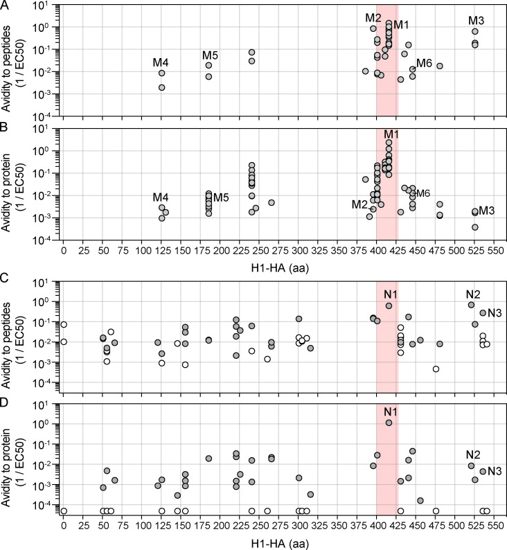
Figure S3.
Functional avidities for peptide and naturally processed H1-HA of T cell clones specific for different epitopes. (A–D) Functional avidity of H1-HA–reactive T cell clones isolated from the memory (A and B) or the naive (C and D) compartment of donor HD1 was determined by stimulation with titrated doses of synthetic peptides (A, n = 37 clones; C, n = 63) or recombinant H1-HA (B, n = 79; D, n = 63). Data are expressed as reciprocal EC50 values. Each dot represents an individual T cell clone; the position of the dots on the x axis indicates the starting residue of the cognate peptide. EC50 values below the detection limit for stimulations with recombinant H1-HA were set arbitrarily to 20 µg/ml; the corresponding T cell clones are reported as white dots. The immunodominant H1-HA401–430 region identified in the memory compartment is highlighted with a red shadow.

