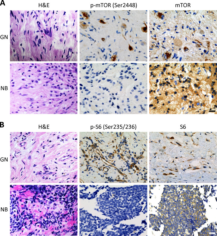
Figure S1.
Immunohistochemical evaluation of mTOR and S6 proteins phosphorylation in human primary ganglioneuromas and poorly differentiated neuroblastomas. (A) H&E staining and IHC with phosphorylated mTOR (p-mTOR; Ser2448) and mTOR for a representative human primary ganglioneuroma (GN) and poorly differentiated neuroblastoma (NB). (B) H&E staining and IHC with phosphorylated S6 ribosomal protein (p-S6; Ser235/236) and S6 for a representative GN and NB. All scale bars represent 20 µm. Protein expression is indicated by brown staining.

