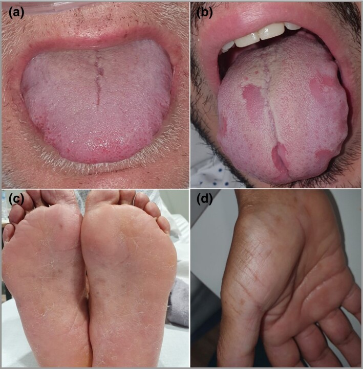
Figure 1.

Upper panel shows COVID‐19 oral mucosa findings. (a) Glossitis with lateral indentations and anterior transient lingual papillitis due to swelling of the tongue and friction with the teeth. (b) Glossitis with patchy depapillation. Lower panel shows palmoplantar findings in patients with COVID‐19. (c) Reddish‐to‐brown acral macules with a slight desquamation on the feet of a patient. Pathology excluded racial pigmentation, showing mild‐to‐moderate lymphocytic infiltrate surrounding the blood vessels and eccrine sweat glands. (d) Acral macules on the palm of a patient with COVID‐19 with the same histopathology.
