Figure 2.

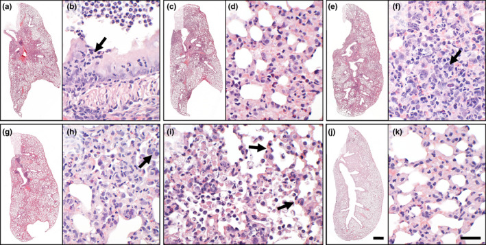
Histopathological assessment of mock‐infected and SARS‐CoV‐2‐infected Chinese hamsters. Histopathology of haematoxylin–eosin‐stained lung sections from SARS‐CoV‐2‐infected Chinese hamsters revealed suppurative bronchitis (a, b; arrow: neutrophils) and necrosuppurative pneumonia (c, d) at 2 and 3 dpi. At 5 dpi (e, f), additional hyperplasia of alveolar epithelial cells (AEC)‐type II was salient (f, arrow). Tissue damage, cell influx and hyperplasia of AEC‐II (h, arrow) were milder but still present at 14 dpi (g, h). Of note, acute alveolar damage (I, arrows) was multifocally distributed throughout the lungs across all time points investigated. None of these lesions was detected in mock‐infected animals (j, k). Bars: 1 mm (a, c, e, g, j) or 50 µm (b, d, f, h, i, k) [Colour figure can be viewed at wileyonlinelibrary.com]
