
Figure 2.
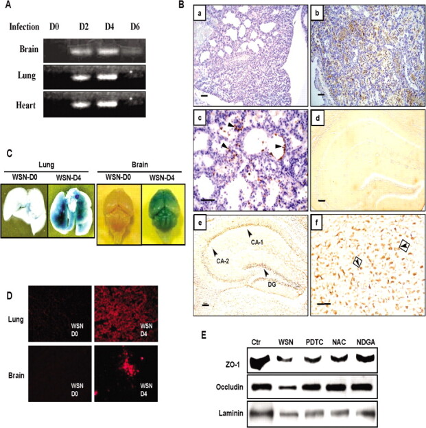
Kinetics of viral proliferation, viral protein accumulation, increase in vascular permeability, and loss of tight-junction proteins in various organs after influenza A WSN/33(H1N1) virus (WSN) infection. A, Detection of viral NS1 gene by reverse-transcription polymerase chain reaction (RTPCR) in the lung, heart, and brain of mice during 0-6 days after infection. B, Immunohistochemical detection of viral antigens in mouse lung and brain at day 4 after infection. a, Hematoxylin and eosin staining of the lung (original magnificationm, ×200). b, Immunoreactive deposits in the lung (original magnification, ×200). c, Viral antigen (arrowheads) in epithelial cells of respiratory bronchioles and infiltrated leukocytes in alveoli (original magnification, ×400). d, No immunoreactive deposits in the brain before infection (original magnification, ×200). e, Virus antigen in the cornu ammonis (CA) 1 and CA-2 and in the stratum granulosum of the dentate gyrus (DG) of the hippocampus (original magnification,×200). f, Virus antigen (arrowheads) in the enlarged image of CA-1 (original magnification, ×400). Scale bars are 100 µm.C, Vascular permeability in the lung and brain analyzed by Evan's blue dye extravasation before (WSN-D0) and after infection at day 4 (WSN-D4). D, Fluorescent micrographs of Evan's blue leakage from capillaries in the brain and lung before and after infection at day 4. E, Loss of tight-junction proteins, zonula occludens (ZO) 1 and occludin, and laminin in the brain analyzed by Western immunoblotting at day 4 after infection and its restoration by pyrrolidine dithiocarbamate (PDTC), N-acetyl-L-cysteine (NAC), and nordihydroguaiaretic acid (NDGA) treatments. The levels before infection are shown as control (Ctr).