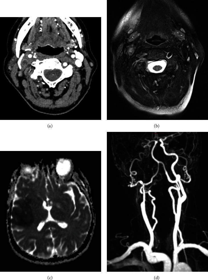
Figure 2.

Axial postcontrast CT image (a) demonstrated the presence of vascular occlusion of the right internal carotid artery, immediately above the bifurcation, than confirmed by T2-weighted turbo spin echo (TSE) axial sequence that suggests also the presence of mural inflammation due to edema detection (b). Axial ADC subtraction sequence at the same time demonstrated the presence of a large frontotemporal ischemic lesion (c). Contrast-enhanced MR-angiography after therapy showed a reduction of the segmental occlusion however with persistent filiform contrasting of the right internal carotid artery (d).
