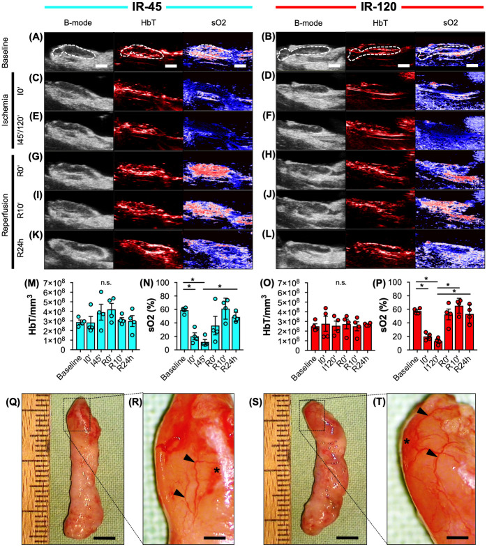
Fig 2. Animal model validation.
A-L Ultrasound (B-mode) and photoacoustic (HbT, sO2) imaging of IR-45 and IR-120 axillary VLN flaps before (A and B), during (C-F) and after (G-L) ischemia (broken line = lymph node). M-P Total hemoglobin (HbT/mm3) and oxygen saturation (sO2 (%)) of IR-45 (M and N, cyan) and IR-120 (O and P, red) lymph nodes at baseline, beginning of ischemia (I0'), after 45 minutes (I45') or 120 minutes (I120') of ischemia as well as directly (R0'), 10 minutes (R10') and 24 hours (R24h) after reperfusion. Mean ± SEM, n = 4, *P < 0.05, n.s. = not significant. Q-T Stereomicroscopic images of IR-45 (Q and R) and IR-120 (S and T) VLN flaps. Higher magnification (R and T = inserts of Q and S) reveals a dilated vascular network (arrowheads) with capsular hemorrhages (asterisks). Scale bars: A-L = 2 mm, Q and S = 4.5 mm, R and T = 900 μm. IR = ischemia/reperfusion, VLN = vascularized lymph node.

