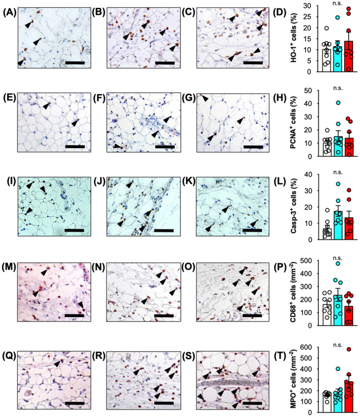
Fig 7. Immunohistochemical analyses of adipose tissue after IR injury.

A-D Immunohistochemical detection of HO-1+ cells (arrowheads) in perinodal adipose tissue of control (A), IR-45 (B) and IR-120 (C) VLN flaps. D Quantitative analysis of HO-1+ cells (%). E-H Immunohistochemical detection of PCNA+ cells (arrowheads) in perinodal adipose tissue of control (E), IR-45 (F) and IR-120 (G) VLN flaps. H Quantitative analysis of PCNA+ cells (%).I-L Immunohistochemical detection of Casp-3+ cells (arrowheads) in perinodal adipose tissue of control (I), IR-45 (J) and IR-120 (K) VLN flaps. L Quantitative analysis of Casp-3+ cells (%). M-P Immunohistochemical detection of CD68+ macrophages (arrowheads) in perinodal adipose tissue of control (M), IR-45 (N) and IR-120 (O) VLN flaps. P Quantitative analysis of CD68+ cells (mm-2). Q-T Immunohistochemical detection of MPO+ neutrophilic granulocytes (arrowheads) in perinodal adipose tissue of control (Q), IR-45 (R) and IR-120 (S) VLN flaps. T Quantitative analysis of MPO+ cells (mm-2). Mean ± SEM, n = 7–8, n.s. = not significant. Scale bars = 50 μm. Casp-3 = cleaved caspase-3, HO = heme oxygenase, IR = ischemia/reperfusion, MPO = myeloperoxidase, PCNA = proliferating cell nuclear antigen, VLN = vascularized lymph node.
