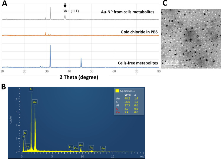
Fig 4. Characterization of AuNPs (AuNP2).
(A) X-ray diffraction (XRD) patterns of the synthesized AuNPs. (B) SEM–EDS (energy dispersive spectroscopy) profile of the synthesized AuNPs. (C) TEM micrographs of AuNPs showed spherical-shaped particles. The samples of AuNPs were placed on standard carbon-coated copper grids (200-mesh) and air-dried for about 2h prior to measurement.

