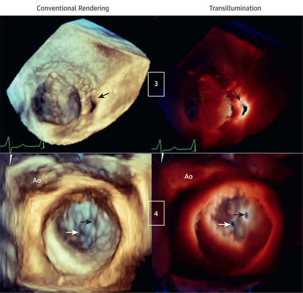
FIGURE 3. Transillumination and Conventional Rendering in Patients With Mechanical Mitral Valve Prosthesis and Perforation of Mitral Valve P3 Scallop.
(Case 3) Bi-leaflet mechanical mitral valve prosthesis with paravalvular leakage (arrows) viewed from the LA. The light is in the LV behind the paravalvular leakage, outlining the orifice edges (Videos 6 and 7). (Case 4) Mitral valve P3 scallop perforation (black arrows) and small, central mal-coaptation (white arrows) are shown. The light is on the valvular plane at the level of the P3 perforation orifice, helping to outline both orifices that otherwise cannot be visualized with conventional rendering (Videos 8 and 9). LV = left ventricle.

