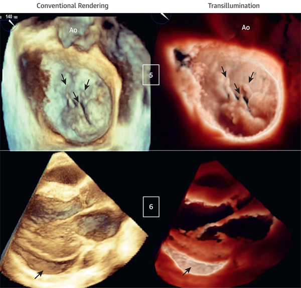
FIGURE 4. Transillumination Versus Conventional Rendering in Patients With Ruptured Mitral Valve Chordae and Pericardial Effusion.
(Case 5) Mitral valve P2 and P3 flail scallops due to multiple ruptured chords (arrows) viewed from the LA. The light is in the LA, close to the valvular plane, slightly anterior to P2. TI rendering enhances the visualization of the ruptured chords and flail scallops (Videos 10 and 11). (Case 6) Inferolateral pericardial effusion (arrows) viewed from a parasternal long-axis acquisition. The light is located deep inside the inferolateral pericardial sac, resulting in a clear definition of the pericardial effusion and cavity (Videos 12 and 13).

