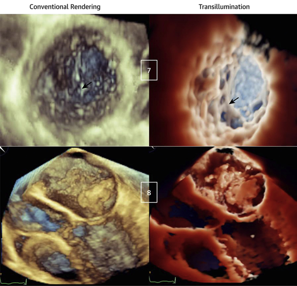
FIGURE 5. Transillumination Versus Conventional Rendering in Patients With Noncompaction Cardiomyopathy (Top) and Left Atrial Myxoma (Bottom).
(Case 7) Apical region of left ventricular noncompaction. The light is in the LV, close to the trabecular inferolateral region, resulting in a clear visualization of the extensive trabeculations in comparison with conventional rendering (arrows) (Videos 14 and 15). (Case 8) Left atrial myxoma with diastolic prolapsing across the mitral valve. The light is in the LA, above the tumor. Although the mass is obvious with conventional rendering, it appears poorly defined, whereas TI rendering underlines mass contours, attachments, dynamic motion, and interaction with the mitral valve (Videos 16 and 17). Abbreviations as in Figures 1 and 3.

