. 2020 Sep 22;9:e55744. doi: 10.7554/eLife.55744

Figure 7. Spermidine and norspermidine restore phagocytic uptake of a spermidine knock-out mutant.

Spermidine was externally added at the indicated concentrations to the ΔPA14_63120 mutant (blue). Using an MOI 1, the phagocytic uptake into RAW264.7 macrophages 1 hr post infection was examined and compared to PA14 and the ΔfliC control. Mean ± standard deviation is displayed. **p<0.01 (one-way analysis of variance (ANOVA), Dunnett’s post-hoc test).
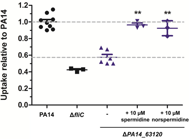
Figure 7.

Figure 7—figure supplement 1. Quantification of spermidine using LC-MS.
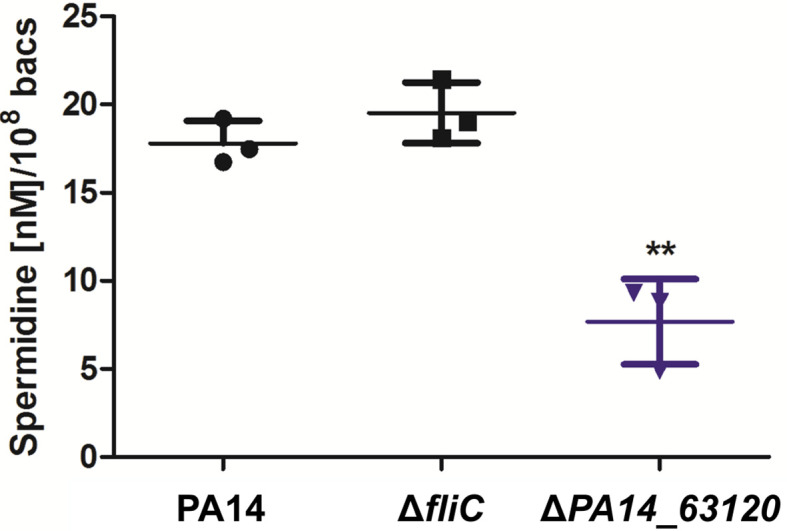
Figure 7—figure supplement 1.

All strains were grown in BM2 media containing glucose as carbon source. Membrane-bound spermidine was extracted from PA14 and its motility variants, derivatized with phenylisothiocyanate and quantified using LC-MS. The amount of spermidine was determined using a calibration curve of adjusted spermidine concentrations and normalized to the bacterial count determined by plating. Mean ± standard deviation of three biological replicates is displayed. **p<0.01 (one-way analysis of variance (ANOVA), Dunnett’s post-hoc test).
Figure 7—figure supplement 2. Effect of norspermidine on phagocytic uptake.
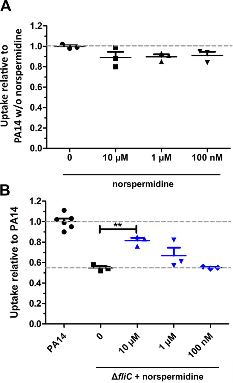
Figure 7—figure supplement 2.

(A) Effect of norspermidine on the phagocytic uptake of PA14 in RAW264.7 macrophages using an MOI of 1. Mean ± standard deviation of three biological replicates is displayed. (B) Effect of 10 µM norspermidine on the phagocytic uptake of ΔfliC on RAW264.7 macrophages using an MOI of 1. Mean ± standard deviation of biological replicates is displayed. **p<0.01 (one-way analysis of variance (ANOVA), Dunnett’s post-hoc test).