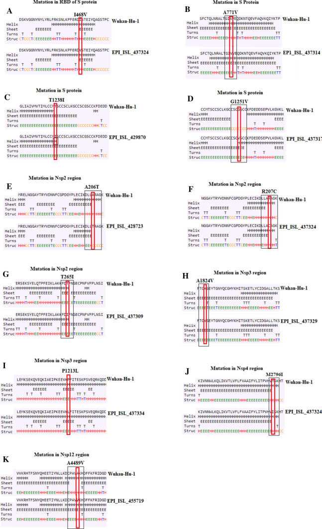
Fig. 1.
Prediction of secondary structure in the S protein, nsp2, nsp3, nsp4, nsp12 regions. a–d Mutations in the S protein. e–g Mutations in nsp2; h–i Mutations in nsp3; j Mutations in nsp4; k Mutations in nsp12. Small rectangular boxes indicate mutated residues Differences in secondary structure between Wuhan and Turkish isolates are indicated by black boxes

