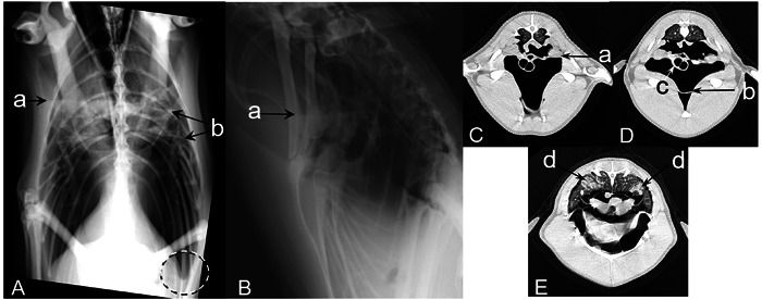
Fig. 1.

Case 1 (King penguin): Radiographs were obtained in the standing position (A and B), computed tomography (CT) were obtained under anesthesia in the supine position (C, D, and E). The fluid level of the clavicular air sac (a), thickening membrane (b), and rounded edge of the air sac (the dashed circle) observed on radiographs and CT images. Thickening of the trachea wall (c) and infiltrate in the pulmonary parenchyma (d) observed only on the CT images.
