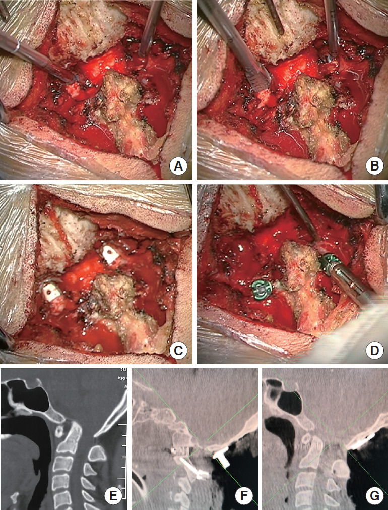
Fig. 3.

Steps in facet joint release and cage implantation in a patient with basilar invagination (BI) and atlantoaxial dislocation (AAD). (A) The joints are bilaterally distracted using a joint loosening tool, and the anterior muscles and ligaments are released. (B) The articular cartilage is widely removed using a joint facet scraper. The size of the spacer depends on the available space within the distracted joint space. (C) Customized lateral mass intervertebral fusion cages, which are tapered at one end for easier placement, are inserted in the joint space. (D) The C2 pedicle screws are placed. Use of the cantilever technique further reduces the horizontal dislocation and adjusts the Oc–C2 angle. (E) A computed tomography (CT) scan shows severe BI-AAD. (F) The intraoperative parasagittal CT image shows the joints with spacers in situ. (G) The intraoperative sagittal CT image shows that the BI and AAD are well reduced.
