Figure 3.
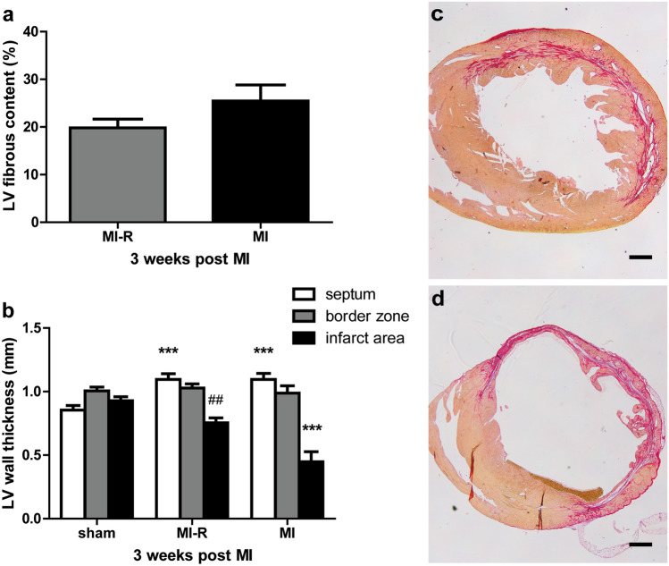
LV fibrous content and wall thickness. Histological analysis after 3 weeks (n = 9–10 per group) showed no significant difference in LV fibrous content between the MI and MI-R group (a). This could possibly be explained since transmural infarction in the MI group caused substantial LV wall thinning which underestimated the fibrous content as a percentage of the total LV when compared to MI-R. LV wall thickness was significantly decreased in the MI group as compared to the MI-R group (b). Sirius red staining of transversal short-axis sections showing typical non-transmural infarction in the MI-R (c) and transmural infarction with substantial LV wall thinning in the MI (d) group. Scale bar: 500 μm. Individual data points are presented in Supplemental Fig. S3. Data are mean ± SEM. ##p < 0.01 versus MI; ***p < 0.001 versus sham.

