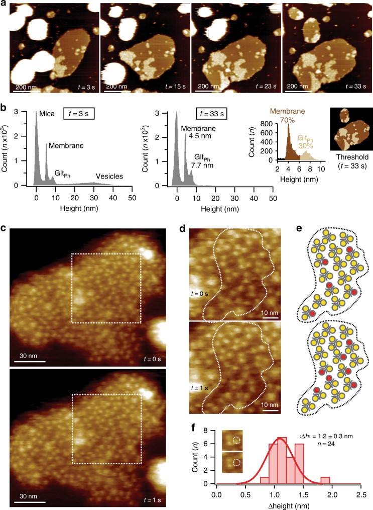
Fig. 1. HS-AFM imaging of GltPh morphology and activity in membranes.
a HS-AFM movie frames of GltPh proteo-liposomes spreading on mica (Supplementary Movies 1 and 2; representative for >50 experimental replicates). b Height distribution analysis: initially (t = 3 s) intact vesicles (height: ~30 nm) adsorb to the surface (defined height: ~0 nm) that eventually (t = 33 s) spread into membrane sheets (height: ~4.5 nm) with GltPh domains (height: ~7.7 nm). Right: membrane packing analysis with thresholds at 3.5 nm < membrane <6.1 nm and 6.1 nm < GltPh < 10 nm. Membranes are ~30% GltPh-packed. c Consecutive high-resolution images. d Zoomed areas of the dashed outlines in c showing GltPh trimers and protomer activity (representative for >50 experimental replicates). e Schematic representation of the dashed outlines in d, where yellow and red circles represent outward- and inward-facing protomers, respectively. f Height change (Δheight) distribution of protomer “elevator” motions in c. Mean ± s.d. are indicated (n = 24). Inset: example protomer (dashed circle).

