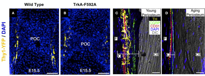
Figure 1.
Nerves in developing, young, and aging bone. (A) Thy1-YFP reporter mice were used to visualize nerve axons in the perichondrial region near the primary ossification center (POC) at embryonic day 15.5. (B) Inhibition of NGF-TrkA signaling using TrkA-F592A mice diminished the density of nerve axons in this region. Scale bars are 100 μm [adapted from (19)]. (C) Utilizing a 120 μm confocal z-stack, CD31+ blood vessels (red), CGRP+ sensory nerve axons (green), and TH+ sympathetic nerve axons (yellow) can be readily visualized in the periosteum of 10-day-old (young) mice. (D) In mice 24 months of age (old), sensory and sympathetic nerve fibers as well as blood vessels remain intact but markedly diminished in the thinner periosteum. Cambium (C) and fibrous (F) layers of the periosteum and cortical bone (CB) are labeled. Scale bars are 15 μm [adapted from (74)].

