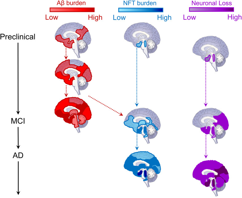
FIGURE 2.
Schematic depiction of the evolution of Alzheimer’s disease biomarkers. Sagittal brain slices depicting the hypothesized locations of Aβ (red, left column) and tau (blue, middle column) deposition, as well as neurodegeneration (neuronal loss, purple, right column) across disease stages. For Aβ, deposition begins in medial prefrontal, cingulate, temporal, and precuneus regions, and progresses cortically and then subcortically until most of the brain contains amyloid plaques (Braak and Braak, 1991; Thal et al., 2002; Sepulcre et al., 2013; Palmqvist et al., 2017). The cortex is likely already saturated with amyloid plaques by the time most patients convert to MCI (Jack et al., 2010, 2013). For tau neurofibrillary tangles (NFT), deposition begins in the locus coeruleus and cholinergic brainstem nuclei in Braak stage 0, progressing into entorhinal cortex in the medial temporal lobe by stage I (Braak and Braak, 1991; Mesulam et al., 2004; Grudzien et al., 2007; Ehrenberg et al., 2017). NFTs remains largely constrained within the MTL until Braak stage III (Braak and Braak, 1991, 1995), when, through its interaction with Aβ pathology (red dashed arrow), tau pathology spreads throughout the brainstem, thalamus, MTL, inferior temporal cortex, and medial prefrontal and cingulate brain regions (Vogel et al., 2020). It is at this time that MCI conversion begins to be observed (Jack et al., 2010, 2013). In later Braak stages, tau pathology is observed throughout most of the cortex (Braak and Braak, 1991, 1995), triggering widespread cortical degeneration as its spreads (Jack et al., 2010, 2013). Regarding neurodegeneration (neuronal loss), as with NFTs, synaptic and neuronal loss begins within locus coeruleus, cholinergic brainstem nuclei, and the MTL (Mesulam et al., 2004; Grudzien et al., 2007; Whitwell, 2010). As MCI progresses into later stages, cortical atrophy is prevalent throughout the temporal, parietal, and occipital lobes (Whitwell, 2010). Ultimately, in later AD stages, cortical atrophy progresses through lateral frontal cortex and, eventually, throughout the entire brain, impacting neural regulation of even basic functions supporting life (Jack et al., 2010, 2013; Whitwell, 2010).

