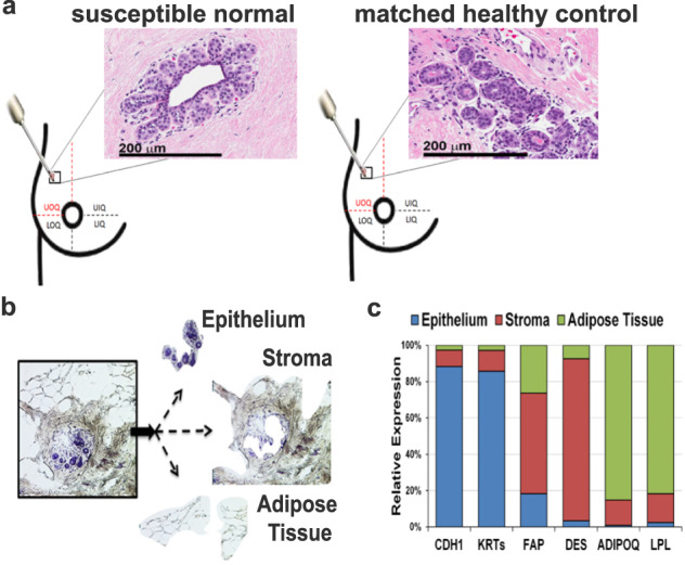
Fig. 2. Microdissection of breast tissue compartments.

a The breast tissue biopsies from women either susceptible to cancer (Susc) or matched healthy controls (HC) were collected from the upper-outer quadrant of the breast (either right or left). Hematoxylin and eosin staining of susceptible and healthy breast tissues showed the lack of any detectable histological breast abnormality at the time of donation. Images at ×40 magnification are shown. Scale bar: 200 µm. b Laser microdissection microscopy was used to isolate the epithelial, stroma, and adipose tissue compartments separately from each of the 23 fresh-frozen biopsies. c Expression of markers specific for the epithelium (E-Cadherin, CDH1, and pan-Keratins (KRTs)), stroma (fibroblast activation protein, FAP, and desmin, DES), adipose tissue (adiponectin, ADIPOQ, and lipoprotein lipase, LPL) was obtained from the transcriptome profiling. Each marker was highly expressed in the specific microdissected tissue compartment as compared with the other areas of the breast (P < 0.0001), thus indicating sample purity.
