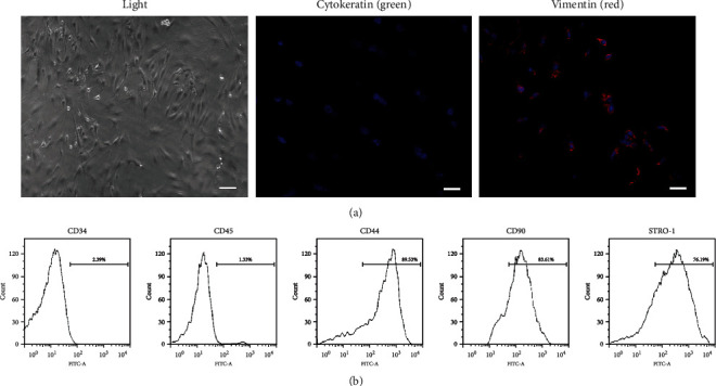
Figure 1.

Characterization of DPSCs by (a) immunofluorescence staining and (b) flow cytometry assay. (a) DPSCs were stained with vimentin (red) and cytokeratin (green). Nucleus was stained by DAPI (blue). (b) DPSCs were stained with endothelial-specific markers (CD34 and CD45) and mesenchymal-specific markers (CD44, CD90, and STRO-1).
