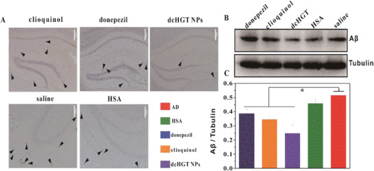
Figure 9.

dcHGT NPs reduced Aβ deposition. A) Immuno‐histochemical staining of Aβ deposition in brain sections of the saline, donepezil, clioquinol, HSA, and dcHGT NP groups. Scale bar: 200 µm. Brown spots are pointed out by arrowheads in the hippocampus of AD mice. B) Western blot analysis and C) quantification of Aβ levels in five groups, with tubulin as a loading control. Data are presented as the mean ± SD. *P < 0.05.
