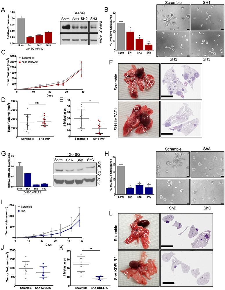
Figure 4. IMPAD1 or KDELR2 expression is necessary for invasive and metastatic ability of lung cancer cells.
a. RT-qPCR and western blot analysis for mouse IMPAD1 expression in 344SQ cells with stable knockdown by shRNA compared to scramble control. b. IMPAD1 knockdown cells form significantly less invasive structures compared to scramble in 3D matrix comprising of 1.5 mg/ml Collagen in Matrigel by day 6 (scale bar: 100uM). See also Supplemental Fig. S6. c-d. Primary tumor growth for 344SQ scramble and SH1 IMPAD1 knockdown cells implanted subcutaneously into syngeneic mice (c) over time, and (d) at time of euthanasia. N=10. e. IMPAD1 knockdown cells form significantly less lung metastatic nodules compared to scramble control. f. Representative lungs and their respective H&E stained sections showing decreased metastases in lungs from mice implanted with SH1 IMPAD1 cells compared to control (scale bar 5mM). g. qPCR and western blot analysis for mouse KDELR2 expression in 344SQ cells with stable knockdown by shRNA compared to scramble control. h. KDELR2 knockdown cells form significantly less invasive structures compared to scramble in 3D matrix comprising of 1.5 mg/ml Collagen in Matrigel by day 6 (scale bar: 100uM). i-j. Primary tumor growth for 344SQ scramble and ShA KDELR2 knockdown cells implanted subcutaneously into syngeneic mice (i) over time, and (j) at time of euthanasia. N=8. k. KDELR2 knockdown cells form significantly less lung metastatic nodules compared to scramble control. l. Representative lungs and their respective H&E stained sections showing decreased metastases in lungs from mice implanted with ShA KDELR2 cells compared to GFP control (scale bar: 5mM). See also Supplemental Fig. S7. Data are represented as mean ± SEM. Significance by Student’s T-test. P-value<0.05 - *; <0.002 - **

