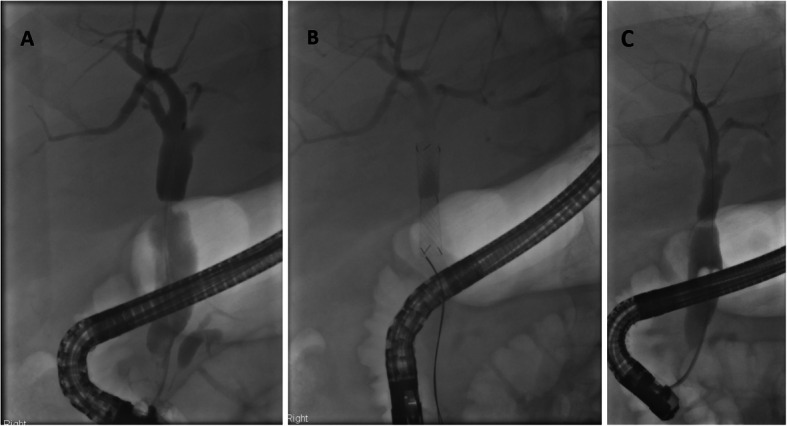
Fig. 3.

The cholangiograms are of a patient who underwent liver transplantation in 2014 for HCC with underling NASH cirrhosis (DBD graft, CIT 8 h). 31 months later they developed cholestatic liver enzymes and imaging showed an anastomotic stricture. They underwent an ERCP and plastic stent insertion but the stent migrated. At the 2nd ERCP (Fig. 3a), the cholangiogram demonstrated the persistence of this stricture. The stricture was dilated using an 8 mm Hurricane Balloon (Boston Scientific) and an 8x40mm Kaffes Stent inserted (Fig. 3b). 3 radiopaque markers on either side of the stent confirm the stent’s position – the top markers represent where the stent begins to be deployed and the middle markers where it should sit over the stricture. Biochemistry immediately improved. The stent was removed 63 days later with complete resolution of the stricture as shown (Fig. 3c)
