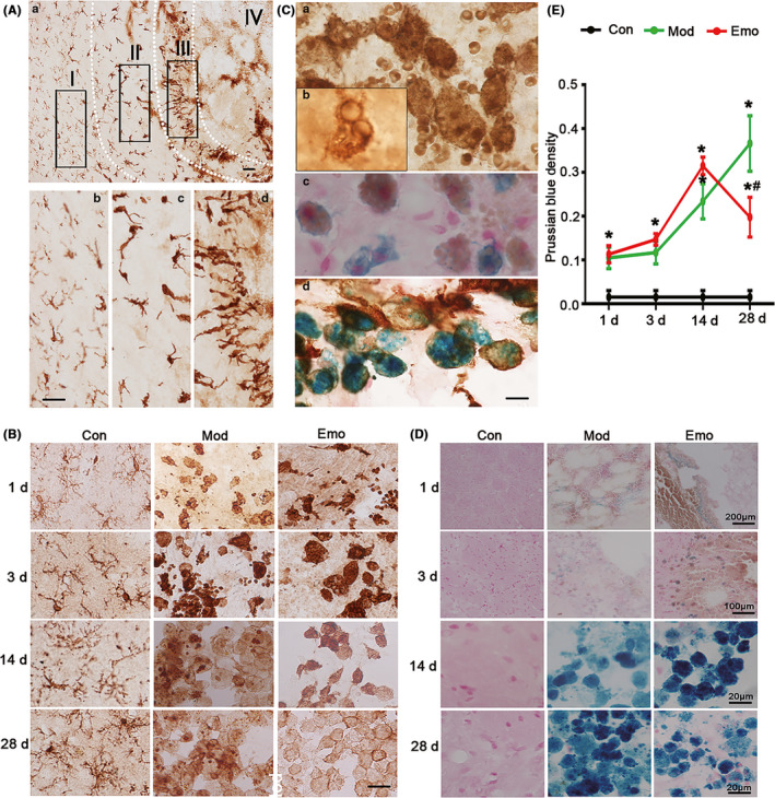
FIGURE 5.

Emodin could improve the morphologic change of microglia/macrophages and reverse the pathological changes of iron deposition in the hemorrhagic CPu of ICH rats. A, After the behavior test, the brain slices (20 μm) in the hemorrhagic CPu were used to assess the morphologic alterations of microglia/macrophages by immunohistochemistry staining and pathological changes of iron deposition by Prussian blue staining. By immunohistochemistry staining on brain slices, Iba‐1 (marker of microglia/macrophage)–positive cells shown different morphological distribution in different areas when collagenase VII was injected in CPu for 3 d. B, Furthermore, Iba‐1–positive cells presented pathological changes in different time and emodin could ameliorate the changes. C, Microglia/macrophages could phagocytize red blood cells which also were positive by Prussian blue staining. D, And then Prussian blue staining was used to value the iron deposition of hemorrhagic lessons. The average degree of density means of Prussian blue stain was detected by IPP software when the injection of 1 d, 3 d, 7 d, 14 d and 28 d. The square area (S) and average degree of mean optical density (MOD, deducting background absorbance) were counted with IPP software and then calculated the density (D, D = S×MOD) of every colored area. E, The total colored square area (ΣS) and total Prussian blue density (ΣD) of every brain section were counted and then the average MOD of total colored area of every brain section (MOD = ΣD/ΣS) was also counted. The data were expressed as means ± SD (n = 10). *P < .05 vs Con, #P < .05 vs Mod
