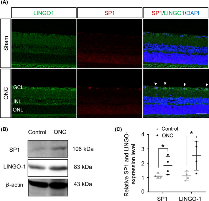
FIGURE 3.

Expression of LINGO‐1 and SP1 was increased in sham and ONC‐injured retinas. A, Representative immunofluorescence staining of LINGO‐1 and SP1 in sham and ONC retinas at 8 d post‐ONC. Scale bar, 50 μm. GCL, ganglion cell layer; INL, inner nuclear layer; and ONL, outer nuclear layer. B, Immunoblotting of SP1 and LINGO‐1 in retinas at 8 d post‐ONC. C, Densitometric analyses of immunoblots of SP1 and LINGO‐1 (n = 4, means ± SD, compared to control retina by Student's t test, *P < .05)
