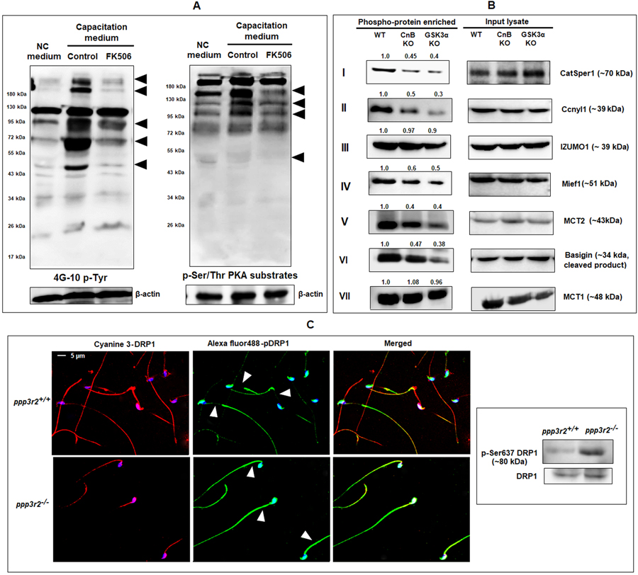
Figure 9: Role of calcineurin in murine sperm protein phosphorylation.
A) During capacitation: Both tyrosine phosphorylated proteins and PKA phosphorylated serine/threonine containing protein profiles were altered under influence of FK506 (20 nM) after 90 min incubation as evidenced by the result of western blot using the anti-mouse 4G10 and anti-rabbit phospho-(ser/thr) PKA substrate antibodies. B), C) Post germ cell differentiation: B) Phosphorylation status of proteins shown were studied by phosphoprotein enrichment analysis. Soluble protein factions extracted from WT, ppp3r2 (CnB) and GSK3a knockout mouse spermatozoa were subject to phospho-enrichment as described under materials and methods. The phospho-enriched fractions and lysates were subjected to western blot analysis with the indicated antibodies. CatSper1, Ccnyl1, Mief1, MCT2 & basigin showed reduced phosphorylation in both CnB and GSK3α knock sperm, while that of IZUMO1 and MCT1 were unchanged (as indicated by the calculated average band intensities provided on top of the corresponding blots). C) Effect of ppp3r2 gene deletion on phosphorylation status of mitochondrial fusion marker DRP1 was determined by immune-cytofluorescence (left panel) and western blot of mouse testis extract (right panel).

