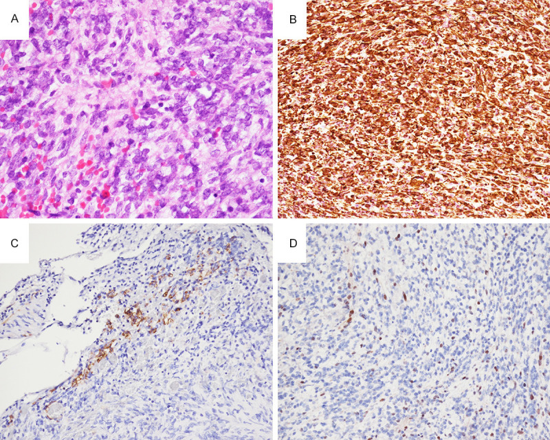
Figure 2.

(A) Hematoxylin and eosin stain of the tumor showed primitive small round to rhabdoid cells with abundant eosinophilic cytoplasm, vesicular chromatin, and conspicuous eosinophilic nucleoli. Tumor cells strongly express vimentin (B), and focally express smooth muscle actin (C). (D) Loss of expression of INI1/SMARCB1 in nuclei of tumor cells, but with retained expression in intratumoral lymphocytes and endothelial cells of blood vessel (200 ×).
