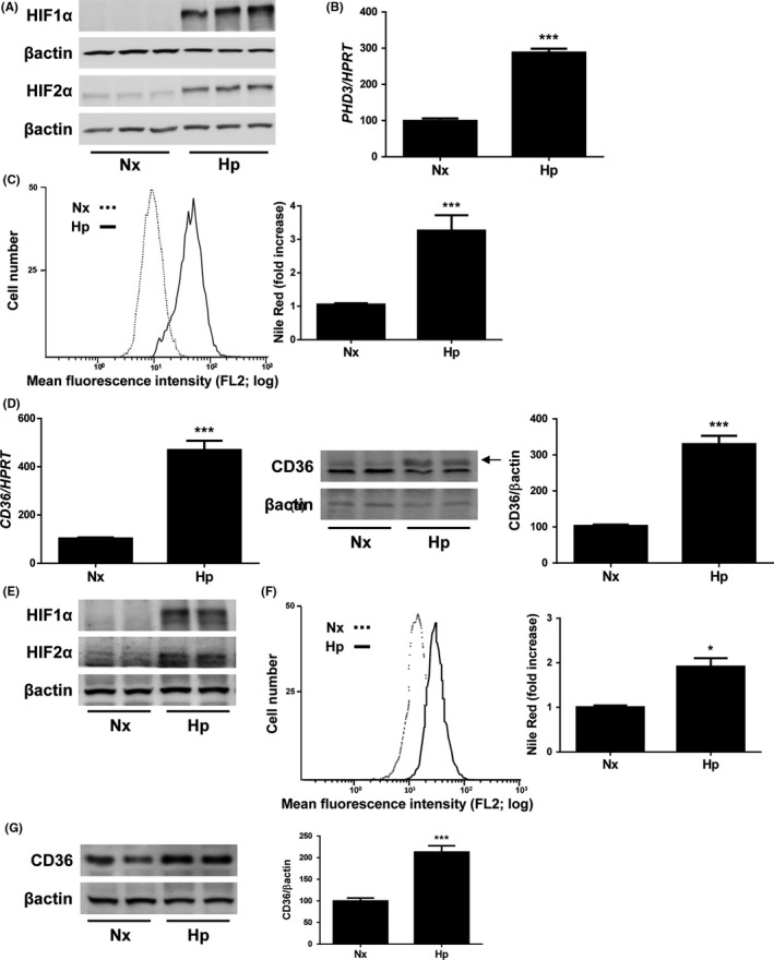
Figure 1.

Hypoxia induces lipid accumulation and CD36 expression in hepatocytes. Huh7 cells maintained under normoxic (Nx, 21% O2), or hypoxic conditions (Hp, 1% O2) in a hypoxia chamber for 36h. A, Representative blots with the indicated antibodies. B, PHD3 mRNA levels. C, (left panel) Representative experiment of Nile Red fluorescence intensity. (right panel) Analysis of intracellular lipid content by Nile Red staining. D, (left panel) CD36 mRNA. (right panel) Representative blots with the indicated antibodies and densitometric analysis from all blots. ***P < .005, Hp vs Nx (n = 4 independent experiments performed by triplicate). AML12 cells maintained under normoxic (Nx, 21% O2), or hypoxic conditions (Hp, 1% O2) in a hypoxia chamber for 36 hours. E, Representative blots with the indicated antibodies. F, (left panel) Representative experiment of Nile Red fluorescence intensity. (right panel) Analysis of intracellular lipid content by Nile Red staining. G, Representative blots with the indicated antibodies and densitometric analysis from all blots. *P < .005 and ***P < .005, Hp vs Nx (n = 3 independent experiments performed by duplicate)
