Fig. 2.

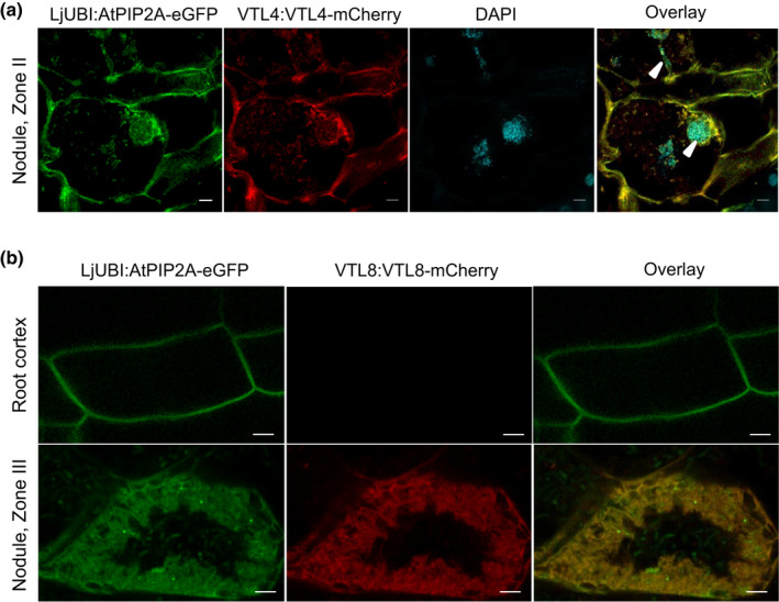
Localization of VTL4 and VTL8 in Medicago truncatula. Translational fusions of VTL4 (a) and VTL8 (b) with mCherry fluorescent protein were expressed in roots of wild‐type seedlings, which were nodulated with Sinorhizobium meliloti 1021. The VTL genes were placed under the control of their own promoters, and co‐expressed with the membrane marker AtPIP2A‐eGFP using the constitutive Lotus japonicus UBIQUITIN (UBI) promoter. (a) Localization of VTL4‐mCherry. The image is of cells in Zone II (infection zone). DAPI staining visualizes DNA including the bacteria in infection threads (arrow heads). Bars, 5 µm. (b) Localization of VTL8‐mCherry. Images of fluorescence signals in the root cortex and Zone III (nitrogen‐fixing zone) are shown. Bars, 5 µm.
