Fig. 8.

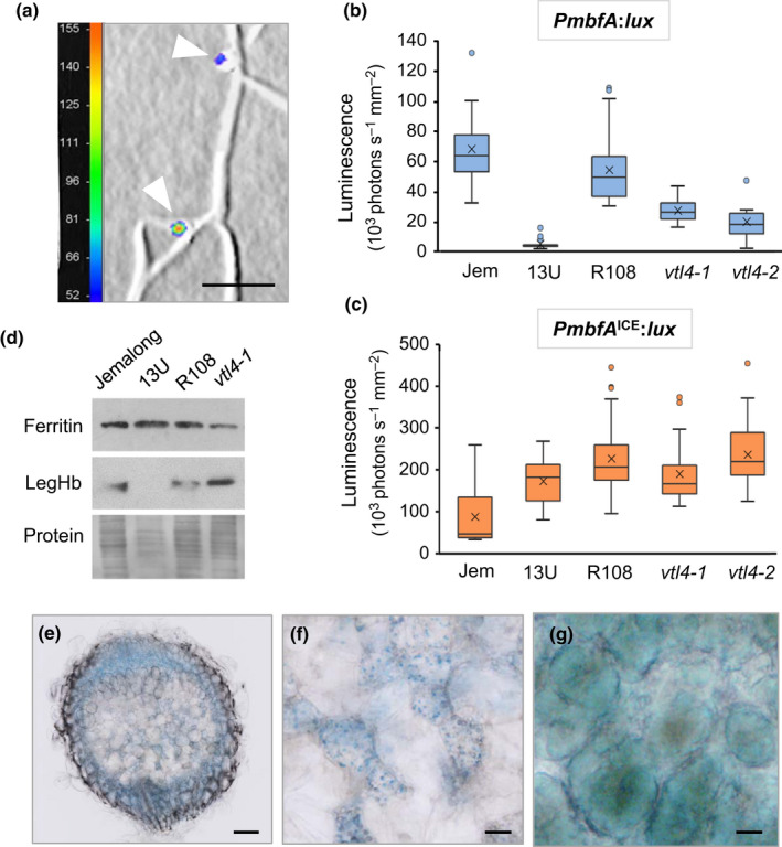
Symbiotic bacteria are iron deficient in nodules lacking VTL4 and VTL8 despite normal iron levels in plant tissues. (a) Detail of an Medicago truncatula R108 root inoculated with Sinorhizobium meliloti 1021 expressing PmbfA:lux at 21 d post inoculation (dpi). Luminescence is represented by a colour scale in photons per second, superimposed on a grey‐scale image. The white arrow heads point to nodules. Bars, 5 mm. (b, c) Expression of the bacterial iron reporter PmbfA:lux (b) and the deregulated Iron Control Element (ICE) mutant (c) in root nodules at 21 dpi, measured as luminescence with a NightOWL camera. Medicago truncatula Jemalong (Jem) was used as wild‐type for the 13U mutant, and M. truncatula R108 as wild‐type for the vtl4 mutants. The data are presented in a box plot, with the box marking the upper and lower quartiles, the middle line represents the median and the symbol × indicates the mean. The whiskers show the spread of the data within the 1.5‐fold interquartile range (n = 9 plants for vtl4‐2, and n ≥ 13 for other lines). (d) Protein blot analysis of ferritin and leghaemoglobin (LegHb) in nodules (21 dpi) of the indicated genotypes. Ponceau S staining of the blot was used as protein loading control (lower panel). (e–g) Histological iron staining of 28‐dpi nodules from (e) 13U mutant plant, (f) enlarged image of the senesced Zone III in 13U, and (g) infected cells in Zone III of wild‐type Jemalong, see Supporting Information Fig. S6 for an image of a whole nodule. Haem‐iron in (g) has a different hue from non‐haem iron. Bars, 0.1 mm (e) and 20 µm (f, g).
