
Figure 1.
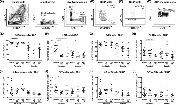
CD4+ memory cells in the decidua parietalis and the decidua basalis. Representative dot plots showing flow cytometric analysis of decidua parietalis live CD4+ lymphocytes using forward/sideward (FSC/SSC) scatterplots, viability stain and CD4 and CD8 expression (A). Within the CD4+ cell population, CD4+ central‐memory (CM) cells (CCR7+CD45RO+CD4+) (B), CD4+ effector‐memory (EM) cells (CCR7−CD45RO+CD4+) (B), general CD4+ memory cells (CM + EM; CD45RO+CD4+) (B), and CD4+ tissue‐resident memory (TRM) cells (CD103+CD45RO+CD4+) (C) were identified. Within the general CD4+ memory cell population, T regulatory (Treg) CD4+ memory cells (Foxp3+CD45RO+CD4+) were identified (D). Proportions of general CD4+ memory cells (E), CD4+ CM cells (F), CD4+ EM cells (G), CD4+ TRM cells (H), Treg CD4+ memory cells (I), Treg CD4+ CM (Foxp3+CCR7+CD45RO+CD4+) cells (J), Treg CD4+ EM (Foxp3+CCR7−CD45RO+CD4+) cells (K), and Treg CD4+ TRM (Foxp3+CD103+CD45RO+CD4+) cells (L), in the decidua parietalis and the decidua basalis from healthy pregnancies and pregnancies complicated by late‐onset preeclampsia (LO‐PE) or early‐onset preeclampsia (EO‐PE), shown as proportion of the CD4+ cell population. Symbols represent individual values per decidua with data as median with interquartile range. Analysis by Kolmogorov‐Smirnov test, one‐way ANOVA and Tukey's post hoc test or Kruskal‐Wallis and Dunn's post hoc test comparing the different groups within the decidua parietalis or the decidua basalis; *P < .05, **P < .01, ****P < .0001