Figure 5.

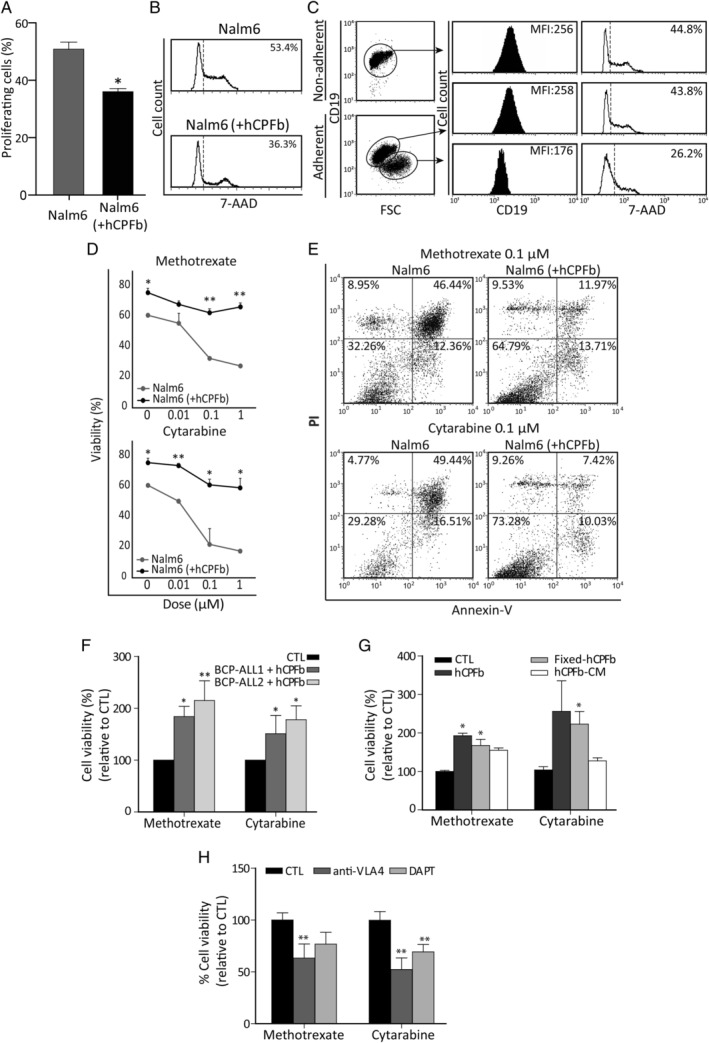
Human choroid plexus fibroblasts protect leukaemia cells from chemotherapy through VCAM‐1/VLA‐4 interactions and Notch signalling. (A) The proliferation rate of Nalm‐6 cells cultured in the presence or absence of human CP fibroblasts (hCPFb) for 48 h was determined by 7‐AAD staining and analysed by flow cytometry. Mean ± SD of proliferating cells from three independent experiments. (B) Representative flow cytometry histograms showing the fraction of cells in S + G2 + M phases. (C) Analysis of leukaemic cell size and CD19 expression when co‐cultured with hCPFb. Dot plots show forward scatter (FSC) properties and CD19 expression of Nalm‐6 cells from non‐adherent (in suspension) and adherent (attached to fibroblasts) fractions. Representative flow cytometry histograms show CD19 expression (solid black histograms) and 7‐AAD staining (open histograms). Mean fluorescence intensity (MFI) and percentage of proliferating cells, respectively, are indicated. (D) The cell viability of Nalm‐6 leukaemic cells exposed to increasing concentrations (0.01, 0.1, and 1 μm) of chemotherapeutic agents (methotrexate and cytarabine) in the presence (black lines) or absence (grey lines) of hCPFb for 72 h was assessed by flow cytometry. Results show the percentage of propidium iodide‐negative and annexin V‐negative viable leukaemic cells, and are the mean ± SD of four independent experiments. (E) Representative dot plots showing the viability of leukaemic cells treated with methotrexate and cytarabine (0.1 μm) in the presence of hCPFb or in control cultures analysed by annexin V and propidium iodide staining. (F) Primary samples from two BCP‐ALL patients (Nos 1 and 2) were exposed to methotrexate and cytarabine (0.1 μm) in the presence or absence of hCPFb. Results from three independent experiments are expressed as the mean ± SD of the relative viability compared with control cultures performed in the absence of hCPFb. (G) Relative viability of Nalm‐6 leukaemic cells co‐cultured with hCPFb, paraformaldehyde‐fixed hCPFb or cultured in the presence of hCPFb‐derived conditioned media and treated with methotrexate and cytarabine at a concentration of 0.1 μm. Data from three independent experiments are expressed as the mean ± SD of the relative viability compared with Nalm‐6 cell control cultures. (H) Cell viability of Nalm‐6 cells treated with chemotherapeutic agents and co‐cultured with hCPFb in the presence of anti‐VLA‐4 blocking antibodies or the Notch inhibitor, DAPT, compared with control cultures (CTL: anti‐IgG or DMSO). Results represent the mean ± SD of three to four independent experiments (*p ≤ 0.05, **p ≤ 0.01; Mann–Whitney test).
