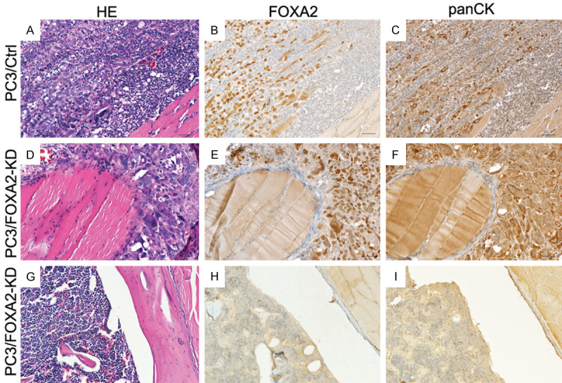
Figure 4.

H&E and immunohistochemical staining of FOXA2 and pan-cytokeratin. (A-C) are serial sections derived from a tibia injected with PC3/Control cells. (D-F) are serial sections derived from a tibia that was injected with PC3/FOXA2-KD cells but demonstrated bone destruction. (G-I) are serial sections derived from a tibia that was injected with PC3/FOXA2-KD cells and did not generate bone destruction.
